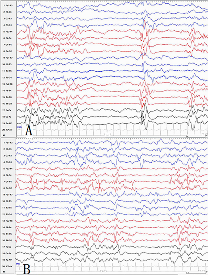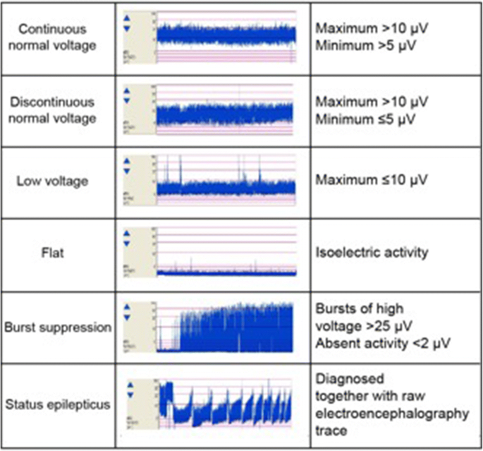
Previous studies also compared the performances by the basic features and segmentation methods. These all represent features employed in the time domain, as well as occasionally the basic features of the frequency domain, like 3 or 10 Hz power or mean power spectral density (PSD). The methods of burst suppression segmentation developed thus far mainly involve detecting burst events by using certain features, such as Shannon entropy, a nonlinear energy operator, line length, a voltage envelope, and variance using recursive-variance estimation.

Thus, researchers initiated an algorithmic approach to burst suppression segmentation from the method used to manually set the threshold in an EEG pattern, evolving into the current methods in use. The current practice employed for this purpose involves a method based on visual detection, being a rough assessment and time-consuming task subjective to the viewers’ interpretation.

The first step in quantifying the depth of burst suppression involves detecting the burst and suppression to distinguish them, thereby called burst suppression segmentation. Accordingly, researchers have developed methods for quantification of burst suppression through calculations of the occupancy ratio of suppression in burst suppression (BSR), analyses using the duration of suppression (interbursts interval, IBI), and quantitation of a burst suppression probability. In addition, a long duration of suppression has also identified its relevance with a worsening prognosis in certain cases (e.g., brain injuries caused by asphyxia), and, further, the progression of burst suppression has provided important prognostic information in previously conducted studies. In the case of a burst suppression caused by anesthesia, the duration of the burst or suppression varies depending on the level of anesthetic concentration, with high levels identifying their relevance to a long duration of suppression. The pattern found from an anesthetized cat’s brain for the first time would accompany the occurrence of a serious reduction in the brain’s activity and metabolic rate, frequently seen from patients with postanoxic encephalopathy or status epilepticus under parenteral benzodiazepine treatments such as midazolam, those under general anesthesia, those with hypothermia, or those in a coma or from neonates. IntroductionĮlectroencephalogram (EEG) burst suppression represents an inactivated EEG pattern, in which the aperiodic alternation of an isoelectric pattern (suppression) and a high voltage pattern (bursts) appears. Burst suppression quantification necessitated precise burst suppression segmentation with an easy optimization therefore, the excellent discrimination and the easy optimization of burst suppression by the proposed method appear to be beneficial. In addition, probabilistic modeling provided a more simplified optimization than conventional methods.

The accuracy was higher than the sole use of the time or frequency domains, as well as conventional methods conducted in the time domain. We evaluated the performance of the proposed method in terms of its accordance with the visual scores and estimation of the burst suppression ratio.

We obtained the feature used in the proposed method from the joint use of the time and frequency domains, and we estimated the decision as to whether the measured EEG was a burst segment or suppression segment by the maximum likelihood estimation. We developed a method to distinguish bursts and suppressions for EEG burst suppression from the treatments of status epilepticus, employing the joint time-frequency domain.


 0 kommentar(er)
